
A glance at any scientific or medical article reveals how far microscopy has evolved from the basic microscope of high school biology classes. Today, it's not just about magnification but also about capturing sharp details, enhancing depth of field, observing internal structures, and experiencing vivid 3D-like visuals that captivate our imagination. Over time, there have been numerous advancements in light microscopy, introducing many new types that unveil a previously unimaginable level of detail.
Stock Image Gallery of Medical Micrographs
The conventional microscope, which is familiar from childhood and known as the "light microscope," functions by utilizing light and a series of lenses. By incorporating filters, specialized mirrors, lasers, specific light spectrums, and other enhancements, we can extract significantly more detailed information.
First, let's cover a few basics:
What is Microscopy

Electron micrograph of red blood cells and fibrin. © Scoatt Camazine / Science Source
Microscopy could be defined as the scientific technique used to observe objects that are too small to be seen with the naked eye, typically using microscopes. It allows researchers to examine the structure and properties of materials and biological specimens at a microscopic level.
Magnification and Resolving Power
Magnification refers to the degree to which an object is enlarged when viewed through a microscope. It is usually expressed as a ratio (e.g., 100x), indicating how many times larger the object appears compared to its actual size.
Resolving power (or resolution) refers to the microscope's ability to distinguish between two closely spaced objects as separate entities. It determines how clearly individual details can be seen.
A Gallery of Royalty-free Micrographs
Let's start with Light Microscopes:

Four types of Light micrographs: Bright Field, Dark Field, Polarized, Phase Contrast. © Marek Mis/Science Source
Various illumination techniques are employed when examining slides under a light microscope, including Bright Field, Dark Field, Fluorescence, Differential Interference Contrast (DIC), Phase Contrast, and Confocal microscopy.
Photos of the microscopes can be found below.
Light Microscope Using Bright Field
Bright Field Microscopy: The light source is directed beneath the specimen, resulting in a brightly lit area surrounding it.
Light Microscope Using Dark Field
Dark Field Microscopy: Here, the light source is obscured, casting light onto the specimen from various angles, providing slightly different details compared to direct illumination.
Fluorescence Microscope
Fluorescence Microscopy: This technique utilizes light filters and specific wavelengths to induce fluorescence in the specimen, producing vibrant images.

Purkinje neurons of the cerebellar cortex stained with four staining methods: hematoxylin eosin (top left), cresyl violet (top right), Cajal's silver nitrate (bottom left) and silver method for Golgi apparatus (bottom right). © Jose Luis Calvo/Science Source
Phase Contrast Microscope
Phase Contrast Microscopy: By employing specialized lenses and filters, this method allows the visualization of transparent and colorless specimens with minimal shadowing.
Differential Interference Contrast Microscope
Differential Interference Contrast Microscopy (DIC): Through the use of polarizers, beam splitters, condensers, and filters, transparent specimens can be observed with a distinctly three-dimensional appearance.

Different illumination techniques of a light microscope: dark field, fluorescence, bright field, phase contrast, DIC (differential interference contrast). Child's hair strand © Ted Kinsman/Science Source

Caenorhabditis elegans (roundworm) embryo transform into a Dauer state when stressed during larval development (for instance, due to lack of food, hot environment, or crowding). Somatic cells are colored red, and yellow dots show gut granules used for fat storage. Image compiled from scans of different layers of the embryo. © Kul Bhatia / Ming Kin Wong / Science Source
Confocal Microscope
Confocal Microscopy: Utilizing Confocal Laser Scanning Microscopy (CLSM), a laser and spatial pinhole combine to produce high-resolution images with enhanced clarity.
Advanced types of microscopes:

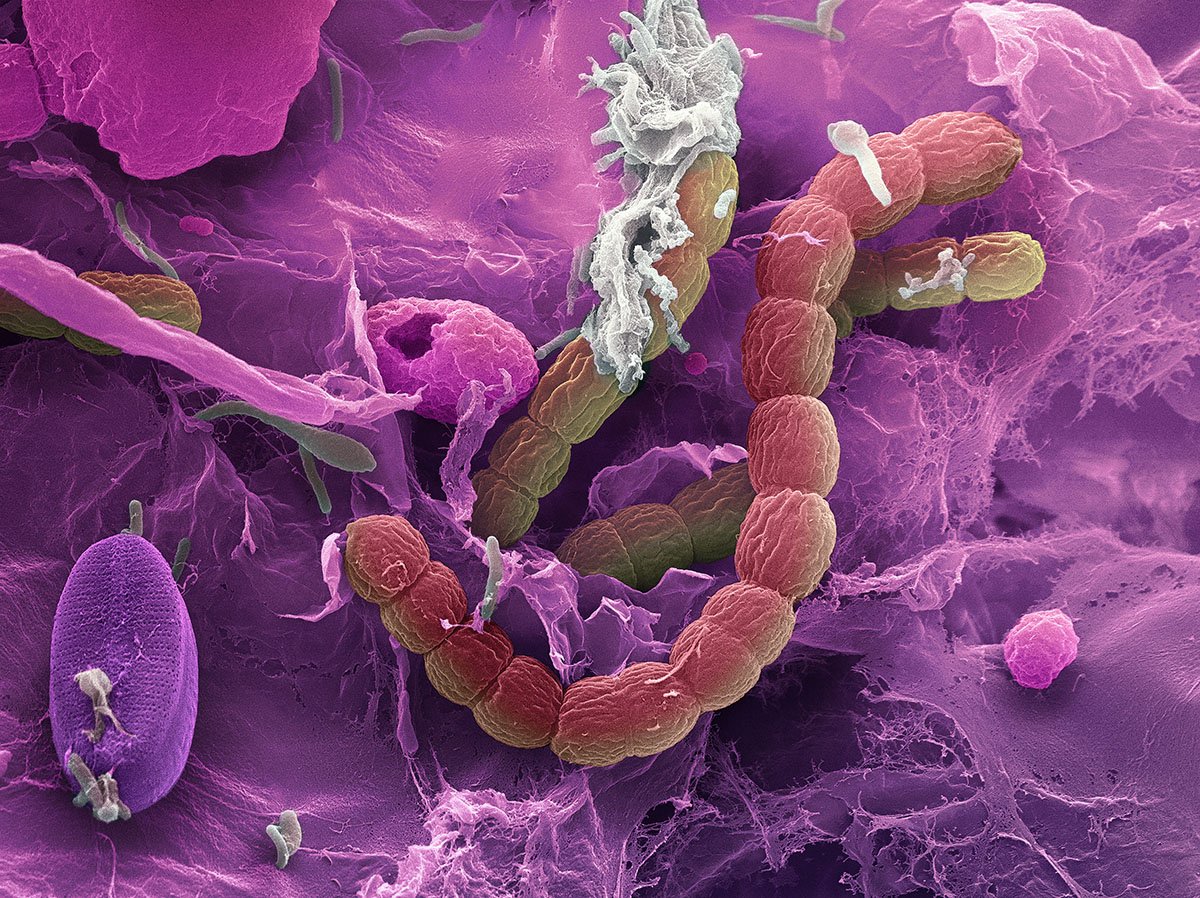
Scanning electron micrograph (SEM) of Escherichia coli (E. coli), a type of Gram-negative rod-shaped bacteria that normally lives in the intestines of people and animals. Magnification is 17,500x at 15cm image size. ©Nano Creative / Science Source
Scanning Electron Microscope
Scanning Electron Microscopy (SEM): This microscope utilizes a beam of electrons to generate sharply detailed images by detecting reflected electrons off the specimen's surface within a vacuum environment, offering magnifications ranging from 20x to 30,000x with a spatial resolution of 50 - 100nm.

Transmission electron micrograph (TEM) of bacteriophage. © Biophoto Associates / Science Source
Transmission Electron Microscope
Transmission Electron Microscopy (TEM): By passing a beam of electrons through a thinly sliced specimen, TEM reveals intricate internal structures with magnifications of up to 2,000,000x.

Plasma membrane proteins. Colored atomic force micrograph (AFM) showing the surface of a plasma membrane. The membrane consists of a lipid bilayer with many proteins (peaks) attached to it. © PROF. DR. H.OBERLEITHNER, UNIVERSITY HOSPITAL OF MUENSTER / Science Source
Atomic Force Microscope
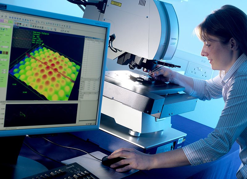
Atomic Force Microscopy (AFM): AFM operates by bouncing a laser off a stylus attached to a cantilever lever, which then traces the specimen's surface. Any deviations trigger sensors, generating a raster image that includes the Z-Plane. Unlike electron microscopy, AFM doesn't require a vacuum, offering versatility in various environments.

False-color scanning tunneling micrograph (STM) of a DNA double-helix; approximately three turns of the helix are displayed in this image. © Driscoll, Youngquist, & Baldeschwieler / Caltech / Science Source
Scanning Tunneling Microscope
Scanning Tunneling Microscopy (STM): STM employs electrons based on quantum tunneling, allowing for atomic-level surface imaging in vacuum, air, water, or ambient gas environments.
What can we look at with all of these microscopes?
Scanning Electron Micrograph (SEM) of unidentified freshwater bacteria collected from pond water. © Ted Kinsman / Science Source
Scoop up pond water or ocean water to be astonished by the plethora of living zooplankton and phytoplankton visible within a single drop using a simple light microscope.
Gallery of Phytoplankton and Zooplankton
It opens you to the wonder of cyanobacteria, blue-green algae, and ciliates like paramecium, daphnia, amoebas, and euglena. If you were lucky, you might have witnessed them conjugate and divide!

Diatom (Stephanopyxis sp.) from a South Atlantic Ocean deep sea probe. Scanning electron microscope. © Eye of Science / Science Source
Additionally, ocean water drops allow a peek at copepods, immature mollusks, krill, algae, crustaceans, and fish in their zooplankton stage. You may also behold the breathtaking beauty of diatoms, the most common type of phytoplankton in our oceans.
Switch to a higher powered Scanning Electron Microscope (SEM) to view Water Bears, pollen, blood cells, and insects. An SEM uses a particle beam of electrons to photograph the surface of a vacuum-sealed specimen.
Transmission Electron Microscopes (TEM) allow us to see cross-sections of specimens, such as the beautiful interior of the human body, marine life, and animal and plant cells. The TEM's particle beam passes through its vacuum-sealed specimen.
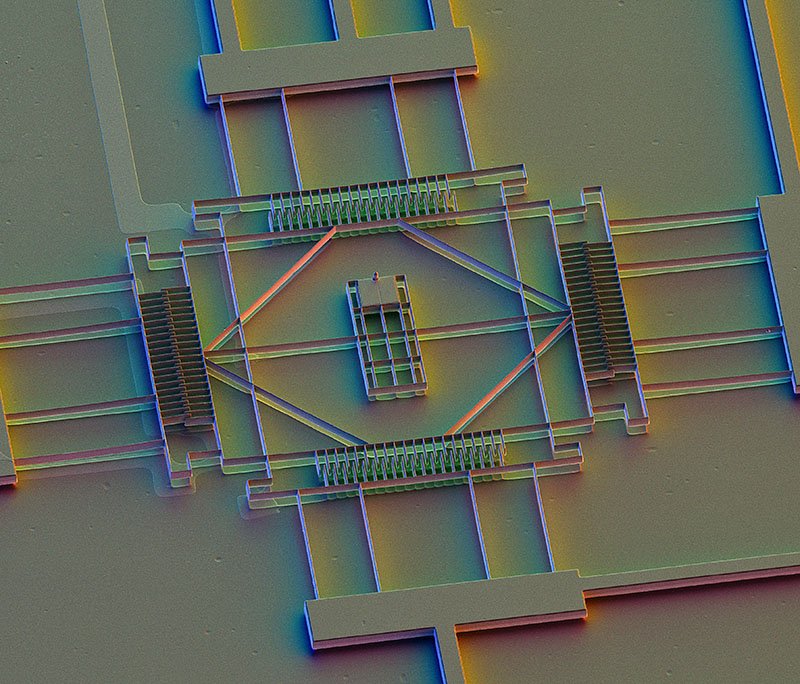
Photos of Types of Microscopes More Photos of Microscopes


Applications of medical microscopy

Bone marrow cell. This micrograph shows this cell with light (inset) and electron microscope. © Jose Luis Calvo/Science Source
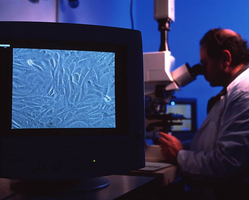
We can view the cells of the human body with many different microscopes. The light mic, SEM, and TEM show scientists and medical researchers different angles and aspects of the cell and its fine structures and organelles.
Microscopes help scientists study cancer - breast, ovarian, prostate, liver, and skin cancer. We can better understand skin conditions such as psoriasis and eczema. They assist in the fight against nervous, respiratory, and circulatory system diseases. And a cure for muscular conditions such as fibromyalgia and multiple sclerosis (MS), and autoimmune conditions.
The Future of Microscopy:
Microscopy methods continue to evolve, with innovations like Acoustic Microscopy, which utilizes sound waves akin to SONAR, promising exciting prospects for the future of scientific exploration.