
Antonie van Leeuwenhoek was born in 1632 to a basket maker and the daughter of a brewer. Though he never received a formal scientific education, he went on to change the future of medicine, biology, chemistry, ecology, and many other fields that rely on magnification as a critical tool.
After apprenticing at a linen-draper's shop in Amsterdam, van Leeuwenhoek moved to Hypolytusbuurt and opened his own business selling fabrics and ribbons to seamstresses and tailors. Always seeking improvement and naturally curious, he wanted a way to examine fabric more closely to assess its quality.
At the time, magnifying glasses were widely available and typically produced by hand-grinding glass. Van Leeuwenhoek took a different approach—he melted a tiny piece of soda-lime glass, forming a small yet exceptionally high-quality lens. The result was a success.
His interest in optics quickly expanded beyond textiles. He crafted increasingly powerful lenses and began observing a growing list of objects: blood cells, skin, rainwater, pond water, plants, mold, insects, and even lice. Most remarkably, he became the first person to observe microscopic organisms, which he famously called "animalcules."
Word of his discoveries eventually reached the Royal Society of London, where his work was met with both skepticism and astonishment. After repeated verification of his observations, he became recognized as the "Father of Microbiology."
The Evolution of Microscopy
Van Leeuwenhoek’s handcrafted lenses paved the way for modern microscopes. Today, scientists use a variety of microscopes, including:
Light Microscope – Uses visible light to magnify objects, commonly used in biology and medicine.
Transmission Electron Microscope (TEM) – Uses electron beams to create highly detailed images of thin specimen slices.
Scanning Electron Microscope (SEM) – Produces three-dimensional images by scanning the surface of a specimen with an electron beam.
Fluorescence Microscope – Uses fluorescent dyes and specialized light to highlight specific cell structures.
Confocal Microscope – Creates high-resolution, 3D images by scanning a specimen layer by layer.
Van Leeuwenhoek could never have imagined the advanced microscopes of today, but his relentless curiosity and ingenuity laid the foundation for a field that continues to revolutionize science.
You can find every type of microscope image for your stock content needs at Science Source. We are known for our live and helpful customer service. Please send us a no-obligation email with any questions.

Leeuwenhoek Microscope
Leeuwenhoek microscope in use. Antonie Philips van Leeuwenhoek (1632 - 1723) is best known for his work on improving the microscope and his contributions towards establishing microbiology. © Biophoto Associates / Science Source
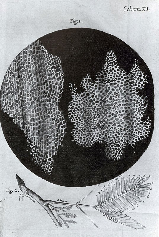
Hooke’s Cork Wood Cells, Illustration
Illustration of cork wood cells by Robert Hooke from observations of cork wood under a microscope. The illustration appeared in his book "Micrographia," which was published in 1667. © Omikron / Science Source

Santiago Ramon y Cajal Looking Through a Microscope.
Portrait of Santiago Ramon y Cajal (1852-1934). Spanish Neuroscientist. © Album / Oronoz / Science Source

Robert Hooke's Louse
An illustration of a louse by Robert Hooke, published in his Micrographia (1665).
Researcher Using a Transmission Electron Microscope
Transmission electron microscopy, 1960s. Researcher using a transmission electron microscope.